
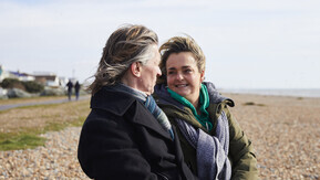
Cancer information and support
If you or someone you care about has been diagnosed with cancer, we're here to help. Use our search to find support and information about cancer.
Cancer information and support
If you or someone you care about has been diagnosed with cancer, we're here to help. Use our search to find support and information about cancer.
Cancer A to Z
Find information about all types of cancer, including diagnosis, treatments and drugs, as well as advice to help with the different ways cancer may impact your life.

Find the right information for you
Answer a few quick questions and we’ll show you the support that fits your situation.
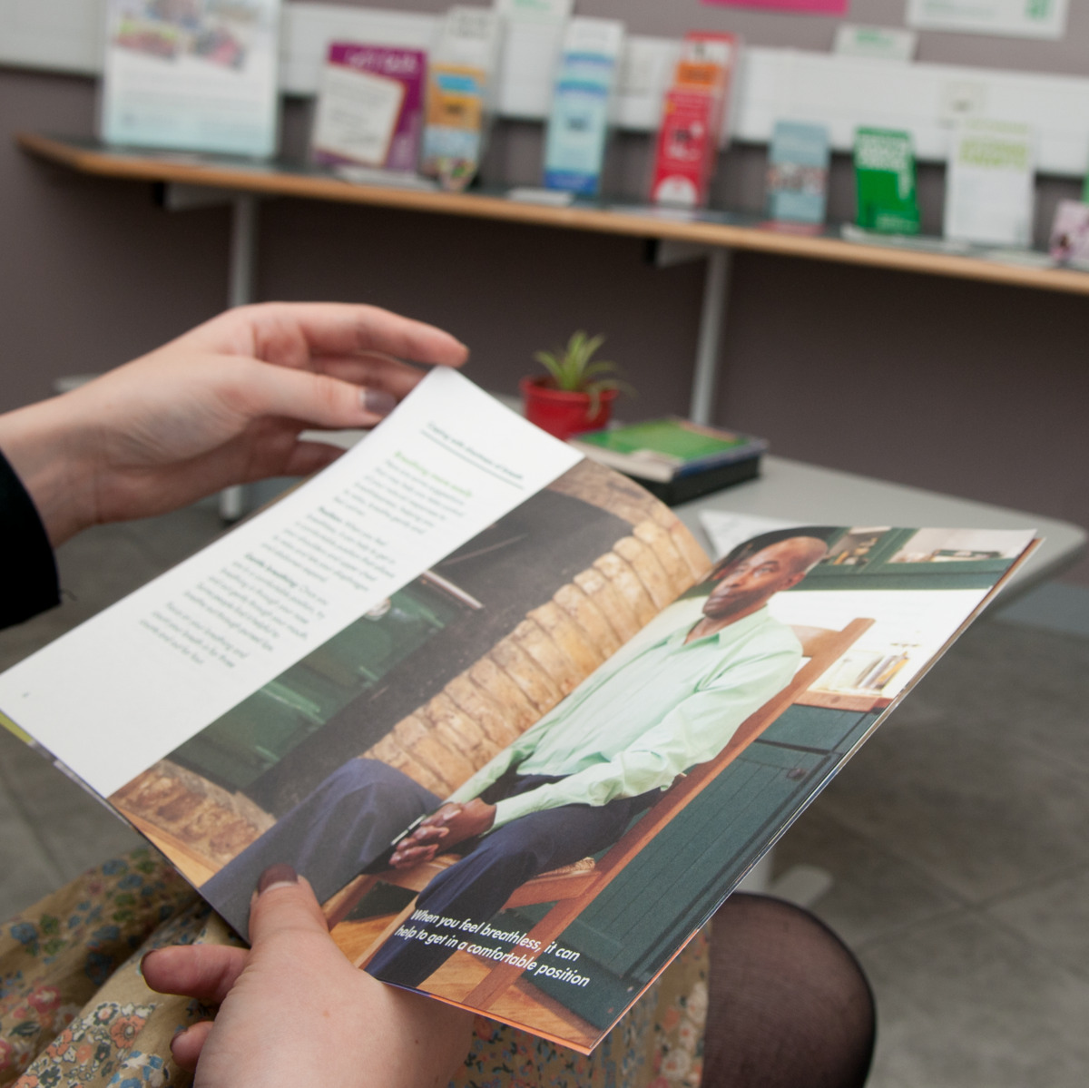
Learn about cancer
Find information about getting a diagnosis, what to expect from treatment, and managing practical and financial worries.


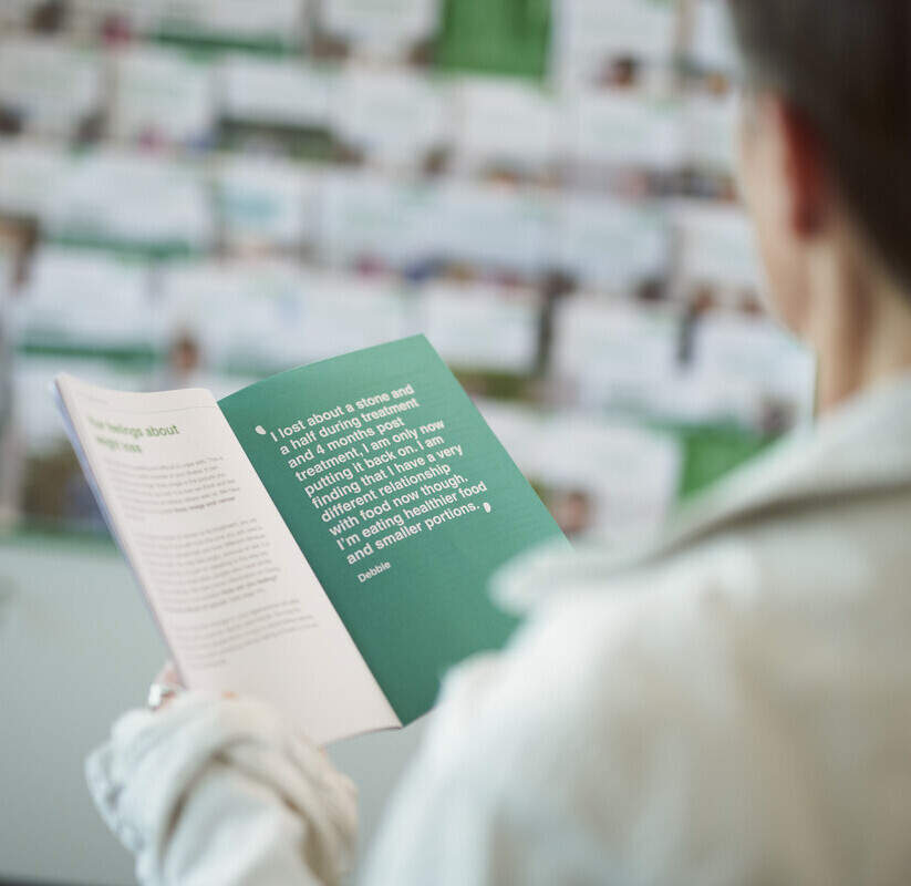
Get information for you
Our information covers all aspects of a cancer diagnosis, and it can be tricky to know where to start. Find the right information for you.
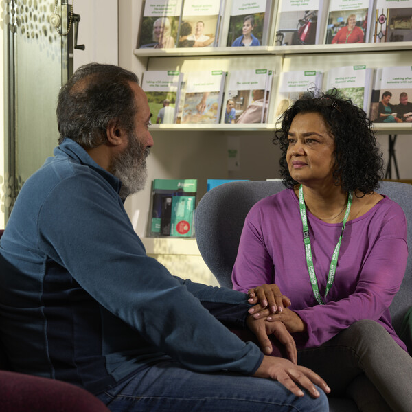

Find cancer support services near you

Which location are you interested in?
Find out about Macmillan services in your area. From cancer information centres to welfare and benefits advice services.