Ultrasound scan
What is an ultrasound scan?
An ultrasound scan uses sound waves to build up a picture of the internal organs. It can also be used to guide a a biopsy
Depending on the type of ultrasound you are having, it may be done by a doctor (radiologist) or a radiographer who is specially trained to do ultrasound scans (sonographer). They are also sometimes done by specialist nurses.
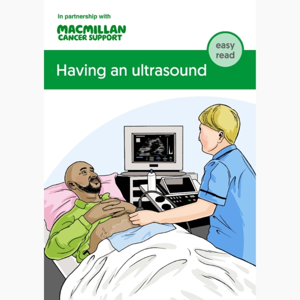
The person doing the scan uses a small device called an ultrasound probe. The probe produces sound waves. These bounce off different parts of the body and make echoes. A computer converts the echoes into a picture.
Depending on the part of the body being scanned, the person doing the scan either:
- moves the probe over the skin (external ultrasound)
- gently inserts it into an opening in the body (internal ultrasound).
Booklets and resources
Having an ultrasound
Depending on the part of the body being scanned, you may be asked to either:
- not eat or drink for a few hours before the scan
- drink plenty of water but not go to the toilet until after the scan.
You may also be asked to change into a hospital gown.
Types of ultrasound
There are 3 main types of ultrasound scan. You can read more about the types of ultrasound scan in the information about the type of cancer you may have.
External ultrasound
This type of ultrasound looks at organs and tissues that can be checked through the skin. These include the liver, kidneys, muscles and joints.
Once you are lying comfortably on your back, the person doing the scan spreads gel on the skin, over the area being scanned. They pass the probe over the area. The scan is painless and takes between 15 and 45 minutes.
Internal ultrasound
There are 2 types of internal ultrasound:
-
Transvaginal ultrasound
Transvaginal ultrasound looks at the ovaries and womb. You lie on your back. The person doing the scan passes a small, lubricated probe gently into the vagina.
-
Transrectal ultrasound
Transrectal ultrasound looks at the back passage (rectum) and prostate. You lie on your left side with your knees bent up. The person doing the scan passes a small, lubricated probe gently into the rectum.
Internal ultrasound scans are not usually painful. But they may be uncomfortable. They usually take 10 to 15 minutes.
Endoscopic ultrasound (EUS)
For an endoscopic ultrasound (EUS), the probe is attached to a thin, flexible tube called an endoscope. A doctor or nurse inserts the tube into the body.
You may have an EUS to look at:
- the digestive system, such as the gullet (oesophagus), stomach and pancreas.
- the throat and lungs. If you have it for the throat and lungs, it is called an endobronchial ultrasound (EBUS).
You usually have a sedative to help you relax. Sometimes you have a general anaesthetic.
For the ultrasound, you lie on your side. The doctor passes the endoscope into your mouth and through to the area being looked at.
An endoscopic ultrasound takes between 15 and 45 minutes. It can be uncomfortable, but it is not usually painful. It may make you feel sick.
After the ultrasound
After the scan is finished, you can usually go home. If you had a sedative, you should:
- not drive for 24 hours
- have someone to take you home
- have someone to stay with you overnight.
If you have had an anaesthetic, you may have to stay in hospital for a few hours.
Date reviewed
This content is currently being reviewed. New information will be coming soon.


Our cancer information meets the PIF TICK quality mark.
This means it is easy to use, up-to-date and based on the latest evidence. Learn more about how we produce our information.